Saturday, May 7, 2016

CONTENTS


General Human Biology & Disease:- 
Elementary Physics and Chemistry.
Characteristics of living matter.
The Structure of living matter
The Tissues
Systems and various parts of human body.
Development and types of Bones.
Bones of Head & trunk
Bones of the Limb
Joints and Articulations
Structure and Action of muscles
The Chief Muscles of the Body
The Blood
The heart and Blood Vessels
The Circulatory system
The Respiratory System
The Digestive System
The Liver, Biliary system and -------
Nutrition and Metabolism
Endocrine Glands and Exec-------
The Urinary System
The Nervous System
The Ear.
The Eye
The Skin
The Reproductive System etc.

Dental Anatomy & Physiology Pharmacology, Pathology including Microbiology.

General & Dental Anatomy. 
Elementary Knowledge of the jaws and teeth, Important between deciduous and permanent teeth. Chronology of eruption , elementary knowledge of occlusion of teeth. Relationship of teeth with investing tissues, muscles of mastication facial expressions and elementary knowledge of temporary mandibular joints.

General & Dental Physiology and Histology. 
Elementary knowledge of the structure and function of various dental and oral tissues e.g. gingival, periodontal membranes, alveollan process, cementum, enamel, dentine, mucous-membranes , pulp.
Salivary glands and functions of saliva, mastication.

General and Dental Pharmacology. 
The therapeutics of drugs commonly used in dentistry and their effects. Practical diagnosis, dispensing of drugs.

Dental Radiology 
Technical aspect of Dental Radiograph i.e. the taking processing and mounting of Dental Radiographs, Radiation Hazards and protection against radiation.

GENERAL DENTISTRY INCLUDING ORAL SURGERY.
Chair side Assistants: -
Reception of Patient.
Lay –out of reception room and Dental Surgery and Hygienist Clinic.
Chair side Assistance and Techniques
Local anaesthesia and equipment.
Methods of Sterilization and care of Dental Instruments.
Basic Principles in surgery
The use of instruments in Dental practice.
Examination of Oral Cavity and Charting of teeth etc.
Instructions to patients and recalls.
Maintenance of Dental Unit/Instrument.

PROSTHODONTICS WITH COSMETOLOGY. 
Introduction and applied anatomy
History taking and Examination
Simple surgical preparation, impression taking.
Selection of patient.
Phonetics and anatomical articulation.
Clasp retained partial denture-plan, treatment, design and management.
Partially edentulous arches.
Connectors – major and minor and functions.
Retainers direct and indirect
Dentures functions, biomechanics, Survey, diagnosis, planning, partial and temporary relining, resilient lining, aids to retention and relief.
Cosmetology and appearance
Dental Materials and its manipulation.

ORTHODONTICS:- 
Etiology, Classification and malocclusion.
Skeletal maturation, growth, dentition with special reference to endocrines.
Classification of dentofacial abnormalities, anthropometrics, cephalometrics.
Examination of patient, differential diagnosis and treatment planning.
Principle of mechanotherapy.
Basics about tweed method, twin wire appliance, activators, plates appliances including tissue reaction and evaluation of treatment.


GENERAL HYGIENE, NUTRITION, COMMUNITY WELFARE CONSERVATIVE & PREVENTIVE DENTISTRY.

DENTAL HYGIENE AND ORAL PROPHLAXIS 
Definition of Hygiene.
Objective of Dental Hygiene.
Oral prophylaxis-various methods.
Stains on teeth-and their management.
Dental Plague, Dental calculus.
Brief description and the role of oral Prophylaxis in Gingivitis, Peritonitis etc.

CLINICALS:-
Instruments, technique of Oral Prophylaxis.
Polishing of teeth.
Topical application of fluorides.
Care of Oral Cavity and appliances during treatment of Maxillo-facial cases.

CONSERVATIVE & PREVENTIVE SURGERY. 
Dental Caries-Prevalence and Prevention.
Periodontal Diseases, saliva in relation to Dental Health and diseases.
Dental Health &Diseases.
Dietary habits and dental Health, Malocclusion, Oral Cancer.

For any correction / suggestion or complaint please feel free to mail me at ayaanmaqsood@gmail.com or call (+91) 9419026242

Friday, April 29, 2016

OCCLUSION OF TEETH

Occlusion means simply the contact between teeth. More technically, it is the relationship between the maxillary and mandibular teeth when they approach each other, as occurs during chewing or at rest.

The study of occlusion is an important aspect of dentistry. The study and practice of most branches of dentistry should be based on a strong foundation of the knowledge of occlusion. Angle defined occlusion as the normal relation of the occlusal inclined planes of the teeth when the jaws are closed. The definition is an oversimplification of what actually it constitutes. Occlusion is a complex phenomenon involving the teeth, periodontal ligament, the jaws, the temporomandibular joint, the muscles and the nervous system.

TERMINOLOGY:
A number of terms are used in the study of occlusion. The following are the commonly used terms.

  • Ideal Occlusion : It is a preconceived theoretical concept of occlusal, structural and functional relationship that includes idealized principles and characteristics that an occlusion should have.
  • Physiologic Occlusion : This refers to an occlusion that deviates in one or more ways from ideal yet it is well adapted to that particular environment, is esthetic and shows no pathologic manifestations or dysfunction.
  • Balanced Occlusion : An occlusion in which balanced and equal contacts are maintained throughout the entire arch during all excursions of the mandible.
  • Functional Occlusion : It is defined as arrangement of teeth, which will provide the highest efficiency during all the excursive movements of the mandible which are necessary during function.
  • Therapeutic Occlusion : An occlusion that has been modified by appropriate therapeutic modalities in order to change a non-physiologic occlusion to one that is at least physiologic if not ideal.
  • Traumatic Occlusion : Traumatic occlusion is an abnormal occlusal stress, which is capable of producing or has produced an injury to the periodontium.
  • Trauma from Occlusion : It is defined as periodontal tissue injury caused by occlusal forces through abnormal occlusal contacts.

PERIODS OF OCCLUSAL DEVELOPMENT:
Occlusal development can be divided into the following developmental periods:
  1. Pre - dental period.
  2. Deciduous dentition period.
  3. Mixed dentition period.
  4. Permanent dentition period
(1) PRE - DENTAL PERIOD : This is the period after birth during which the neonate does not have any teeth. It usually lasts for 6 months afterbirth.
The alveolar processes at the time of birth are known as gum pads. The gum pads are pink, firm and are covered by a dense layer of fibrous periosteum. They are horse shoe shaped and develop in two parts. They are the labiobuccal portion and the lingual portion. The two portions of the gum pads are separated from each other by a groove called dental groove. The gum pads are divided into ten segments by certain grooves called transverse grooves. Each of these segments consists of one developing deciduous tooth sac.
The gingival groove separates the gum pad from the palate and floor of the mouth. The transverse groove between the canine and first deciduous molar segment is called the lateral sulcus. The lateral sulci are useful in judging the interarch relationship at a very early stage. The lateral sulcus of the mandibular arch is normally more distal to that of the maxillary arch.
The upper and lower gum pads are almost similar to each other. The upper gum pad is both wider as well as longer that the mandibular gum pad. Thus when the upper and lower gum pads are approximated there is a complete overjet all around. Contact occurs between upper and lower gum pads at the first molar region and a space exists between them in the anterior region. This infantile open bite is considered normal and it help in suckling.
The neonate is without teeth for about 6 months of life. At birth the gum pads are not sufficiently wide to accommodate the developing incisors which are crowded in their crypts. During the first year of life the gum pads grow rapidly permitting the incisors to erupt in good alignment.
Very rarely teeth are found to have erupted at the time of birth. Such teeth that are present at the time of birth are called natal teeth. Sometimes teeth erupt at an earlu age. Teeth that erupt during the first month of age are called neonatal teeth. The natal and neonatal teeth are mostly located in the mandibular incisor region and show a familial tendency.

(2) DECIDUOUS DENTITION PERIOD : The initiation of primary tooth buds occurs during the first six weeks of intra-uterine life. The primary teeth began to erupt at the age of about 6 months. The eruption of all primary teeth is completed by 2½ to 3½ years of age when the second deciduous molars come into occlusion.
The mandibular central incisors are the first teeth to erupt into the oral cavity. They erupt around 6-7 months of age. (Click here to read chronology of eruption). The average eruption of the deciduous dentition is given in Table 1. The timing of tooth eruption is highly variable. A variation of three months from the mean age has been accepted as normal. The sequence of eruption of the deciduous dentition is :

The primary dentition is usually established by 3 years of age on eruption of the 2nd deciduous molars. Between 3 - 6 years of age the dental arch is relatively stable and a very few changes occur.
Space usually exists between the deciduous teeth. These spaces are called physiological spaces or developmental spaces. The presence of spaces in the primary dentition is important for the normal development of the permanent dentition. Absence of spaces in the primary dentition is an indication that crowding of teeth can occur when the larger permanent teeth erupt.
Spacing invariably occurs mesial to the maxillary canines and distal to the mandibular canines. The physiological spaces are called Primate spaces or Simian spaces as they are seen commonly in primates. These spaces help in placement if the canines of the opposing arch.

Saturday, April 2, 2016

Chronology of Eruption

The axial or occlusal movement of tooth from its developmental position within the jaw to its functional position in the occlusal plane is known as eruption of teeth.


There are three types of movements:

  • Pre - Eruptive
  • Eruptive
  • Post - Eruptive
PRE - ERUPTIVE : When deciduous tooth germ differentiates, there is a good deal of space between them. But due to their rapid growth, this available space is utilized and developing tooth become crowded together, especially in incisor and canine region. This crowding is relieved by growth in length of infant jaws which provides room for second deciduous molars to drift backward and anterior teeth to drift forward. At the same time, the tooth germ also moves outwards as jaw increases in width and height.
Permanent teeth with deciduous predecessors also undergo complete movement before they reach the position from which they erupt. As their deciduous predecessors erupt, they move to a more apical position and occupy their own bony crypt. Premolars begin their development lingual to their predecessors at the level of occlusal surface and in same bony crypt. They are situated beneath the divergent roots of deciduous molars. The permanent molars which do not have predecessors also move from the site of their initial differentiation.
ERUPTIVE : There is axial or occlusal movement of tooth from its developmental position within the jaw to its final functional position in the occlusal plane. It is important to recognize that jaw growth is normally occurring while most teeth are erupting, so movement in plane other than axial is superimposed on eruptive movement.
POST - ERUPTIVE : These movements are those that :-

  • Maintain the position of the erupted tooth which the jaw continues to grow.
  • Compensates for proximal and occlusal wear.


THEORIES OF TOOTH ERUPTION
  1. Bone Re-modelling
  2. Root growth
  3. Vascular pressure
  4. Periodontal ligament traction


(a) BONE RE-MODELLING :- It supposes that selective deposition and resorption of bone brings eruption. In the experiments, where tooth germ is removed and the follicle is left in the position the eruptive pathway still form in bone. Thus this indicates the dental follicle and not bone as major determinant in tooth eruption.

(b) ROOT GROWTH : Root formation is also unlikely to be the cause of tooth eruption; as the onset of root formation is not synchroneous with onset of axial tooth movement. It causes overall increase in length of tooth that must be accompanied by root growing in the bone of jaw by an increase in jaw length or by crowing moving occlusally.
But it is not accepted. For example, if erupting tooth is prevented from erupting by pinning it to the bone, root growth continues and is surrounded by resorption of bone at base of socket.Thus although it can produce force, root growth can not be translated into eruptive movement unless there is some structure at the base of tooth, capable of withstanding this force but no such structure exists.
Advocates of the root growth theory postulated the existence of a ligament, the cushion - hammock ligament, straddling the base of the socket from one bony wall to the other like a sling. But the hammock ligament is the pulp delineating membrane that runs across the apex of the tooth and has no bony insertion so it can not act as a fixed base.

(c) VASCULAR PRESSURE : It states that there is a higher pressure system either within or around the base of tooth. It is known that teeth move in synchrony with arterial pulsation, so local volume changes can produce limited tooth movement.
Whether such pressure is prime for movement of teeth is debatable because surgical excision of the root and therefore the local vasculature does not prevent tooth eruption.

(d) PERIODONTAL LIGAMENT TRACTION : There is good deal of evidence that eruptive force resides in the dental follicle-periodontal ligament complex. As long as periodontal tissue is available tooth movement occurs. Tissue culture experiments have shown that ligament fibroblast is able to contract a collagen-gel which in turn brings about movement of a disc of root tissue attached to that gel. Thus, there is no doubt that periodontal ligament fibroblasts have the ability to contract and transmit a contractile force to the extra cellular environment and in particular to the collagen fiber bundles in vitro. The entire morphological features exist in vivo to permit similar movement.
In summary, eruptive movement is brought about by a combination of events involving a force initiated by the fibroblasts. This force is transmitted to the extra cellular compartment via fibron exuses and to collagen fiber bundles, which aligned is an appropriate inclination brought about by root formation, bring about tooth movement. These fiber bundles must have the ability to remodel for eruption to continue and interference with this ability affects the eruptive process. The removal of bone to create the eruptive pathway is also dictated by the tissue surrounding the tooth.

CHRONOLOGY OF ERUPTION
CHRONOLOGY OF ERUPTION OF PRIMARY TEETH
TEETH
ERUPTION AGE
Mandibular Central Incisor
6 Months
Mandibular Lateral Incisor
7 Months
Maxillary Central Incisor
7 ½ Months
Maxillary Lateral Incisor
9 Months
Mandibular First Molar
12 Months
Maxillary First Molar
14 Months
Mandibular Canine
16 Months
Maxillary Canine
18 Months
Mandibular 2nd Molar
20 Months
Maxillay 2nd Molar
24 Months


CHRONOLOGY OF ERUPTION OF PERMANENT TEETH

Teeth (listed in order of eruption sequence)
Eruption time
Mandibular 1st Molar
6 – 7 Years
Maxillary 1st Molar
6 – 7 Years
Mandibular Central Incisor
6 – 7 Years
Maxillary Central Incisor
7 – 8 Years
Mandibular Lateral Incisor
7 – 8 Years
Maxillary Lateral Incisor
8 – 9 Years
Mandibular Canine
9 – 10 Years
Maxillary 1st Premolar
10 – 11 Years
Mandibular 1st Premolar
10 – 11 Years
Maxillary 2nd Premolar
10 – 12 Years
Mandibular 2nd Premolar
11 – 12 Years
Maxillary Canine
11 – 12 Years
Mandibular 2nd Molar
11 – 12 Years
Maxillary 2nd Molar
12 – 13 Years
Mandibular 3rd Molar
17 – 20 Years
Maxillary 3rd Molar
18 – 20 Years

QUESTIONS:

Q1: How many types of movements are there in the eruption of teeth?
Ans: Three (Pre-Eruptive, Eruptive and Post-Eruptive)

Q2: Which tooth erupts first in the oral cavity?
Ans: Primary Mandibular Central Incisor

Q3: Which permanent tooth erupts first in the oral cavity?
Ans: Mandibular First Molar.

Q3: Which successor tooth erupts first in the oral cavity?
Ans: Mandibular Central Incisor.

Q4: At what age, primary teeth erupt in the oral cavity?
Ans: At the age of 6 months.

Q5: At what age, Primary Mandibular Central Incisors erupt in the oral cavity?
Ans: 6 months

Q6: At what age, Primary Mandibular Lateral Incisors erupt in the oral cavity?
Ans: 7 months

Q7: At what age, Primary Maxillary Central Incisors erupt in the oral cavity?
Ans: 7 1/2 months

Q8: At what age, Primary Maxillary Lateral Incisors erupt in the oral cavity?
Ans: 9 months

Q9: At what age, Primary Mandibular First Molars erupt in the oral cavity?
Ans: 12 months

Q10: At what age, Primary Maxillary First Molars erupt in the oral cavity?
Ans: 14 months

Q11: At what age, Primary Mandibular Canines erupt in the oral cavity?
Ans: 16 months

Q12: At what age, Primary Maxillary Canines erupt in the oral cavity?
Ans: 18 months

Q13: At what age, Primary Mandibular 2nd Molars erupt in the oral cavity?
Ans: 20 months

Q14: At what age, Primary Maxillary 2nd Molars erupt in the oral cavity?
Ans: 24 months

Q15: At the age of 5 months, how many teeth are present in the oral cavity?
Ans: Nill / 0

Q16: At the age of 6 months, how many teeth are present in oral cavity?
Ans: 2 teeth (Mandibular Central Incisors)

Q17: At the age of 8 months, how many teeth are present in oral cavity?
Ans: 6 teeth ( 4 Mandibular Incisors and 2 Maxillary Central Incisors)

Q18: At the age of 10 months, how many teeth are present in oral cavity?
Ans: 8 teeth ( Incisors)

Q19: At the age of 13 months, how many teeth are present in oral cavity?
Ans: 10 teeth ( 8 Incisors and 2 Mandibular First Molars)

Q20: At the age of 15 months, how many teeth are present in oral cavity?
Ans: 12 teeth ( 8 Incisors and 4 First Molars)

Q21: At the age of 17 months, how many teeth are present in oral cavity?
Ans: 14 teeth (8 Incisors, 4 First Molars and 2 Mandibular Canines)

Q22: At the age of 19 months, how many teeth are present in oral cavity?
Ans: 16 teeth (8 Incisors, 4 First Molars and 4 Canines).

Q23: At the age of 21 months, how many teeth are present in oral cavity?
Ans: 18 teeth (8 Incisors, 4 Canines, 4 First Molars and 2 Second Molars)

Q24: At the age of 25 months, how many teeth are present in oral cavity?
Ans: 20 teeth (8 Incisors, 4 Canines and 8 Molars)

Sunday, March 6, 2016

Difference between Deciduous and Permanent Teeth


Humans exhibit two sets of teeth, primary teeth and permanent teeth. Primary dentition erupts first in the oral cavity and is replaced by permanent dentition. The difference between the primary and permanent teeth is given below.

1. Primary teeth are smaller in overall size than the analogous permanent teeth.

2. The colour of the enamel is whiter in primary teeth than the permanent teeth. The tooth colour depends upon the level of mineralization of enamel. The deciduous teeth have less mineralized enamel, which is more opaque and whiter. Highly mineralized enamel is very translucent and the underlying dentin can be seen through the enamel and impart and yellowish tinge to the tooth.

3. The crowns of primary teeth are wider mesiodistally than they are occluso-cervically. Therefore, they are more bulbous than their permanent successors.

4. Primary teeth have a marked cervical prominence, especially on the buccal aspect of primary molars.

5. Primary teeth show a greater degree of attrition than that shown by permanent teeth. Attrition depends upon the level of mineralization of the enamel. More highly mineralized enamel is harder and so is more slowly worn away than less mineralized enamel.

6. Depth of enamel is more constant and thinner in the primary teeth than in permanent teeth.

7. The incisal edges of deciduous incisors show no mamelons.

8. The contact areas between deciduous molars are broader, flater and situated more gingivally than those between permanent molars.

9. The second deciduous molars are decidedly larger than the first molars whereas reverse is true for permanent dentition.

10.  Microscopically, the enamel rods in the gingival one-third of the deciduous molars as they proceed towards the enamel surface incline towards the occlusal, compared to the permanent teeth, where these rods incline cervically.

11. Roots of the primary teeth are longer and thinner mesiodistally in comparison with crown size than those of the permanent.

12. The roots of deciduous teeth are fully formed about one year after eruption whereas the roots of permanent teeth take a longer duration about 3 years.

13. Roots of the primary teeth undergo physiologic resorption, where as in the permanent teeth the resorption is pathologic.

14. Pulp horns of primary teeth are more prominent and are higher in primary molar.

15. Primary pulp ages at a faster rate than the permanent pulp.

16. The radicular pulp of the primary molars follows a thin, tortuous and branching path.

17. There are no clearly defined root canal entrances in primary teeth.

Important Questions for Objective Type Exams:

Q: How many sets of teeth humans exhibit?
Ans: 2

Q: Why primary teeth are more whiter than permanent teeth?
Ans: The deciduous teeth have less mineralized enamel, which is more opaque and whiter.

Q: The tooth colour depends upon the?
Ans: Level of mineralization of the enamel.

Q: Highly mineralized enamel is?
Ans: Very translucent.

Q: Why primary teeth are more bulbous than permanent teeth?
Ans: Because crowns of primary teeth are wider mesiodistally than they are occluso-cervically.

Q: Which set of teeth show greater degree of attrition?
Ans: Primary teeth

Q: Why primary teeth show greater degree of attrition than permanent teeth?
Ans: Attrition depends upon the level of mineralization of the enamel. More highly mineralized enamel is harder and so is more slowly worn away than less mineralized enamel.

Q: Which type of resorption takes places in primary teeth?
Ans: Physiologic resorption.

Q: Which type of resorption takes place in permanent teeth?
Ans: Pathologic resorption.

Q: Which type of pulp ages faster?
Ans: Primary pulp.

Saturday, March 5, 2016

PERMANENT TEETH or ADULT TEETH

Permanent teeth or adult teeth are the second set of teeth formed in diphyodont mammals. In humans, there are thirty-two permanent teeth, consisting of six maxillary and six mandibular molars, four maxillary and four mandibular premolars, two maxillary and two mandibular canines, four maxillary and four mandibular incisors. The first permanent tooth usually appears in the mouth at around six years of age.

The first of the permanent teeth to erupt are the permanent first molars, right behind the last 'milk' molars of the primary dentition. These first permanent molars are important for the correct development of a permanent dentition. Up to the age of thirteen years, twenty-eight of the thirty-two permanent teeth will appear.
The full permanent dentition is completed much later during the permanent dentition period. The four last permanent teeth, the third molars, usually appear between the ages of 17 and 25 years; they are considered wisdom teeth.
It is possible to have extra, or "supernumerary," teeth. This phenomenon is called hyperdontia and is often erroneously referred to as "a third set of teeth." These teeth may erupt into the mouth or remain impacted in the bone. Hyperdontia is often associated with syndromes such as cleft lip and palate, trichorhinophalangeal syndrome, cleidocranial dysplasia, and Gardner's syndrome.

Important Questions for Objective Type Exams:

Q: In diphyodont mammals, the second set of teeth is called?
Ans: Permanent teeth or Adult teeth.

Q: How many permanent teeth are present in humans?
Ans: 32

Q: At what age, the permanent teeth began to erupt in the oral cavity?
Ans: 6 years.

Q: Which set of permanent teeth erupt first in the oral cavity?
Ans: Mandibular first molars.

Q: Which teeth are called as "Wisdom teeth"?
Ans: Third molars

Q: At what age wisdom teeth erupt in the oral cavity?
Ans: 17 - 25 years.

Q: Extra teeth are called?
Ans: Supernumeray teeth

Q: The phenomenon of having supernumeray or extra teeth is called?
Ans: Hyperdontia

Q: The phenomenon of having less number of permanent teeth? or The phenomenon in which permanent teeth are missing?
Ans: Hypodontia

Q: The phenomenon of having no permanent teeth in oral cavity?
Ans: Anodontia

Q: At the age of 13, years how many permanent teeth are present in the oral cavity?
Ans: 28

Friday, March 4, 2016

DECIDUOUS or PRIMARY TEETH

Deciduous teeth, otherwise known as baby teeth, temporary teeth, milk teeth, and now more commonly primary teeth, are the first set of teeth in the growth development of humans and other diphyodont (two sets of teeth) mammals. The word deciduous has come from the Latin word which means to fall off or shed like leaves of a tree. The other terms unfortunately denote a lack of importance because the nick names indicate the usefulness of these teeth for a short period only. They develop during the embryonic stage of development and erupt—that is, they become visible in the mouth—during infancy. They are usually lost and replaced by permanent teeth, but in the absence of permanent replacements, they can remain functional for many years.

Primary teeth start to form during the embryo phase of pregnancy. The development of primary teeth starts at the sixth week of tooth development as the dental lamina. This process starts at the midline and then spreads back into the posterior region. By the time the embryo is eight weeks old, there are ten buds on the upper and lower arches that will eventually become the primary (deciduous) dentition. These teeth will continue to form until they erupt in the mouth. In the primary dentition there are a total of twenty teeth: five per quadrant and ten per arch. The eruption of these teeth ("teething") begins at the age of six months and continues until twenty-five to thirty-three months of age during the primary dentition period. Usually, the first teeth seen in the mouth are the mandibular centrals and the last are the maxillary second molars.

The primary dentition is made up of central incisors, lateral incisors, canines, first molars, and secondary molars; there is one in each quadrant, making a total of four of each tooth. All of these are gradually replaced with a permanent counterpart except for the primary first and second molars; they are replaced by premolars. The replacement of primary teeth begins around age six, when the permanent teeth start to appear in the mouth, resulting in mixed dentition. The erupting permanent teeth cause root resorption, where the permanent teeth push on the roots of the primary teeth, causing the roots to be dissolved by odontoclasts (as well as surrounding alveolar bone by osteoclasts) and become absorbed by the forming permanent teeth. The process of shedding primary teeth and their replacement by permanent teeth is called exfoliation. This may last from age six to age twelve. By age twelve there usually are only permanent teeth remaining.

Primary teeth are essential in the development of the oral cavity. The primary teeth maintain the arch length within the jaw, the bone and the permanent teeth replacements develop from the same tooth germs as the primary teeth. The primary teeth provide guides for the eruption pathway of the permanent teeth. Also the muscles of the jaw and the formation of the jaw bones depend on the primary teeth to maintain proper spacing for permanent teeth. The roots of primary teeth provide an opening for the permanent teeth to erupt. The primary teeth are important for the development of the child's speech, for the child's smile and play a role in chewing of food.


Important QUESTIONS for Objective Type Exams:

Q: A condition where two sets of teeth erupt is called?
Ans: Dipyodonty

Q: A condition where teeth are replaced throughout the life is called?
Ans: Polyphyodonty

Q: How many primary teeth are present in oral cavity?
Ans: 20 teeth (4 quadrants, each quadrant consisting of 1 central incisor, 1 lateral incisor, 1 canine, 1 first molar and 1 2nd molar).

Q: Which set of teeth appear first in the oral cavity?
Ans: Primary or Deciduous or Milk or Baby teeth.

Q: When primary teeth start to form?
Ans: Between sixth and eighth week of prenatal development.

Q: The phenomenon of eruption of primary teeth in the oral cavity is called?
Ans: Teething

Q: At what age, the eruption of the primary teeth begins in the oral cavity?
Ans: 6 - 7 months or 7 - 8 months

Q: At what age, the eruption of all the primary teeth ends?
Ans: 2 ½  to 3 ½  years

Q: Which teeth erupts first in the oral cavity?
(OR)
Which primary teeth erupts first in the oral cavity?
Ans: Deciduous / Mandibular central incisors

Q: Which primary teeth erupt last erupt in the oral cavity?
Ans: Maxillary second molars.

Q: Which set of teeth is absent in primary dentition?
Ans: Premolars

Q: At what age, the replacement of primary teeth begins?
Ans: 6 years of age

Q: The process of shedding primary teeth and their replacement by permanent teeth is called?
Ans: Exfoliation

Q: At what age, all the primary teeth are replaced by the permanent teeth in the oral cavity?
Ans: 12-13 years.

Q: The period when only primary teeth are present in the oral cavity is called?
Ans: Primary Dentition Period

Q: When does the roots of the deciduous teeth begin to resorb?
Ans: 2 - 3 years after their completion.

Q: Exfoliation of the deciduous teeth occurs between?
Ans: 7 to 12 years.

Q: 

Deciduous and Permanent Teeth

Deciduous Teeth : Deciduous teeth, otherwise known as baby teeth, temporary teeth, milk teeth, and now more commonly primary teeth, are the first set of teeth in the growth development of humans and other diphyodont mammals... MORE CLICK HERE

Permanent Teeth : Permanent teeth or adult teeth are the second set of teeth formed in diphyodont mammals... MORE CLICK HERE

Difference between Deciduous and Permanent Teeth : CLICK HERE

Saturday, November 14, 2015

Introduction to Dental Anantomy


The mouth is a versatile area of the human anatomy used for respiratory and digestive functions besides other functions such as speech. Teeth form the most important component of the Oral cavity.
In non-mammalian vertebrates the teeth are constantly replaced throughout life, a condition called Polyphyodonty. Most mammals including humans exhibit two sets of teeth. Usually, at birth no teeth are present in the oral cavity . Soon the primary or deciduous teeth erupt in the oral cavity. These primary teeth are later replaced by permanent teeth. Such a condition where two sets of teeth exist is called diphyodonty.

QUADRANTS OF THE MOUTH:
Oral cavity in humans is divided into four quadrants. Each quadrant resembles other three quadrants in morphology. The upper jaw which is called as maxilla is divided further into two quadrants; (i) right maxillary quadrant and (ii) left maxillary quadrant.Similarly, the lower jaw called as mandible is also divided into two similar quadrants (i) Right mandibular quadrant and (ii) Left mandibular quadrant.


HUMAN DENTAL FORMULA:
The human dentition have teeth of 4 distinct morphological types. They are the incisors, canines, premolars and molars.


The number of teeth present in the deciduous and permanent dentition are different and can be represented by a dental formula. The human dental formula for primary and permanent dentition is given below: The formula represent the teeth of a single quadrant.
Dental Formula for Primary dentition
Dental Formula for Permanent Dentition

The teeth of the upper jaw are called maxillary teeth while the teeth of the lower jaw are called mandibular teeth.
Thus in all there are 20 deciduous teeth and 32 permanent teeth.

THE STRUCTURE OF A TOOTH
Each tooth in humans features two primary components: the crown and the root. 

CROWN : That portion of a tooth which projects above the gum line and is visible in the oral cavity.

ROOT : That portion of a tooth which projects into the jaw base and anchors the tooth.

The crown and the root portion of a tooth meet at cemento-enamel junction.

The crown morphology differs from tooth to tooth, Incisors are flat and possess an incisal edge. Canines have one cusp while premolars and molars have two or more cusps.

INCISORS :
They are named so in man because they are used to incise food. They possess thin blade like crowns which are adapted for cutting and shearing food preparatory to grinding. There are two incisors in each quadrant of the jaw. They are termed the central incisors and the lateral incisors.


CANINES :
The canine is present distal to the incisors. It is a large tooth having a single cusp. Morphologically they can be considered transitional between incisor and the premolar. The canines have the longest root in the jaw.
PREMOLARS :
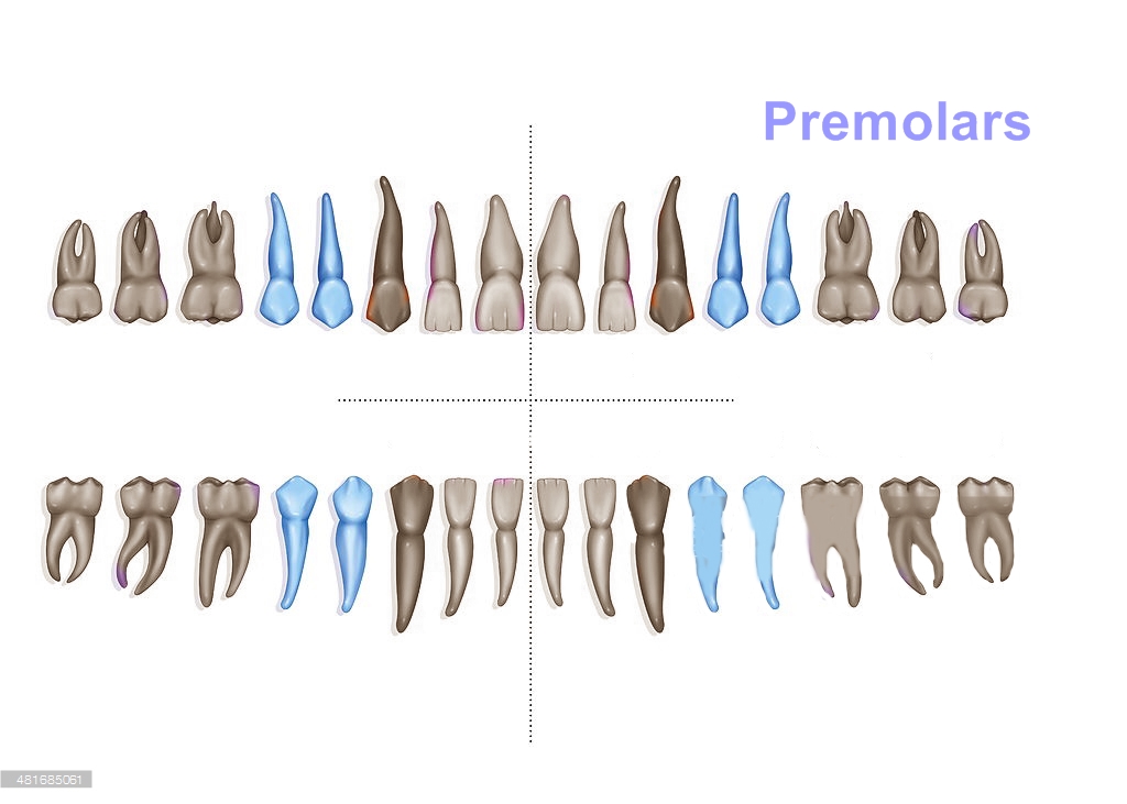
Premolars are found distal to the canine and are unique to the permanent dentition. The replace the deciduous molars. The premolars usually possess two cusps; one buccal and a lingual / palatal, separated from each other by a mesiodistal occlusal fissure. Morphologically the premolars are considered transitional between canines and molars.

MOLARS :
They are teeth that possess the largest occlusal surface having 3 to 5 cusps, The permanent molars do not have deciduous predecessors. They are used for grinding food. The permanent dentition has three molars. They are called the first, second and third molars starting distal of the canine.
The root portion of a tooth can end as a single root or can divide to form two or more roots as seen in molars and certain premolars. The root is held firmly within the jaw base i,e the maxilla and the mandible. The portion of the jaw bone that houses and supports the roots is called the alveolar process.


Monday, June 22, 2015

Characteristics of living matter

All living things, animals and plants, are organisms that undergo chemical processes by which they sustain life and regenerate cells. The difference between animals and plants is that animals have sensations and the power of voluntary movement, and they require oxygen and organic food. On the other hand, plants require only carbon dioxide and inorganic matter for food and have neither voluntary movement nor special sensory organs.

In man, some of the characteristic functions necessary for survival include digestion, metabolism, and homeostasis. Digestion involves the physical and chemical breakdown of the food we eat into its simplest forms. Metabolism is the process of absorption, storage, and use of these foods for body growth, maintenance, and repair. Homeostasis is the body's self-regulated control of its internal environment. It allows the organism to maintain a state of constancy or equilibrium, in spite of vast changes in the external environment.


THE CELL:
The cell, the smallest unit of life, is the basic structural unit of all living things and a functional unit all by itself. Cells are composed of a viscid, jellylike substance, called protoplasm, upon which depend all the vital functions of nutrition, secretion, growth, circulation, reproduction, excitability, and movement. Protoplasm, thus, has often been called "the secret of life."

A typical cell is made up of the plasma membrane, the nucleus, and the cytoplasm.

The plasma membrane is a selectively permeable membrane surrounding the cell. In addition to holding the cell together, the membrane selectively controls the exchange of materials between the cell and its environment by physical and chemical means. Gases (such as oxygen) and solids (such as proteins, carbohydrates, and mineral salts) pass through the plasma membrane by a process known as diffusion.

The nucleus is a small, dense, usually spherical body that controls the chemical reactions occurring in the cell. The substance contained in the nucleus is called nucleoplasm. The nucleus is also important in the cell's reproduction, since genetic information for the cell is stored there. Every human cell contains 46 chromosomes, and each chromosome has thousands of genes that determine the cell's function.

The cytoplasm is a gelatinous substance surrounding the nucleus and is contained by the plasma membrane. The cytoplasm is composed of all of the cell protoplasm except the nucleus.

The simplest living organism consists of a single cell. The amoeba is a unicellular animal. The single cell of such a one-celled organism must be able to carry on all processes necessary for life. This cell is called a simple or undifferentiated cell, one that has not acquired distinguishing characteristics.

In multicellular organisms, cells vary in size, shape, and number of nuclei. Whenstained, the various cell structures can be more readily recognized under a microscope. Other differences such as the number and type of cells can be seen with the aid of a microscope. Many cells are highly specialized. Specialized cells perform special functions (e.g., muscle cells, which contract, and epithelial cells, which protect the skin).

Tuesday, May 5, 2015

Introduction to Dentistry


Dentistry is the branch of medicine that is involved in the study, diagnosis and prevention and treatment of diseases, disorders and conditions of the oral cavity, commonly in the dentition but also the oral mucosa and of adjacent and related structures and tissues, particularly in the maxillofacial (jaw and facial) area. Although primarily associated with the teeth among the general public, the field of dentistry or dental medicine is thus not limited to odontology (from the Ancient Greek 'Odous' means 'tooth') - the study of the structure, development and abnormalities of the teeth.
Because of their substantial overlap in concepts, dentistry is often understood to subsume the now largely defunct, medical specialty of stomatology (the study of mouth and its disorders and diseases) for which the two terms are used interchangeably in certain regions.
Dentistry is widely considered important for overall health. Dental treatment is carried out by the dental team, which often consists of a dentist and dental auxiliaries (Dental Assistants, Dental Hygienists, Dental Technicians and Dental Therapists)
The history of dentistry is almost as ancient as the history of humanity and civilizations, with the earliest evidence dating back from 7000 BCE. Prehistoric dental surgical techniques are seen in Ancient Egypt, where a mandible dated to approximately 2650 BCE shows two perforations just below the root of the first molar, indicating the draining of an abscessed tooth. Remains from the early Harappan periods of the Indus valley civilization (3300 BCE) shows evidence of the teeth having been drilled dating back 9000 years. It is thought that the dental surgery was the first specialization from medicine.
Dr. John M Harris started the world's first Dental school in Brainbridge, Ohio and helped to establish dentistry as a health profession. It opened on Feb 21, 1828 and today is a dental museum. The first dental college Baltimore college of Dental surgery, opened in Baltimore Maryland U.S.A in 1840. The French surgeon Pierre Fauchard became known as the father of the modern dentistry.
Common treatments in dentistry involve the restoration of teeth, extraction or surgical removal of the teeth, scaling and root-planning and endo-dontic root canal treatment. The earliest dental filling, made of beeswax was discovered in Shoveria and dates 6500 years ago.
The two most common dental diseases are dental caries and Pyorrhea.
>>